

Fitness professionals must have a basic understanding of skeletal system anatomy due to the key role it plays in all aspects of human life, including all movement and exercise. The bones, ligaments, tendons, and joints are all key structures in anatomy. Additionally, professionals must understand how disease and resistance training impacts these structures.
Introduction
The skeletal system is an integral part of the human body. It supports body movement and protects vital organs. It serves as an attachment site for muscles and ligaments. In addition, the skeleton stores calcium, which is necessary for many body functions including cardiac function. Bones are also the primary production site for some types of blood cells.1
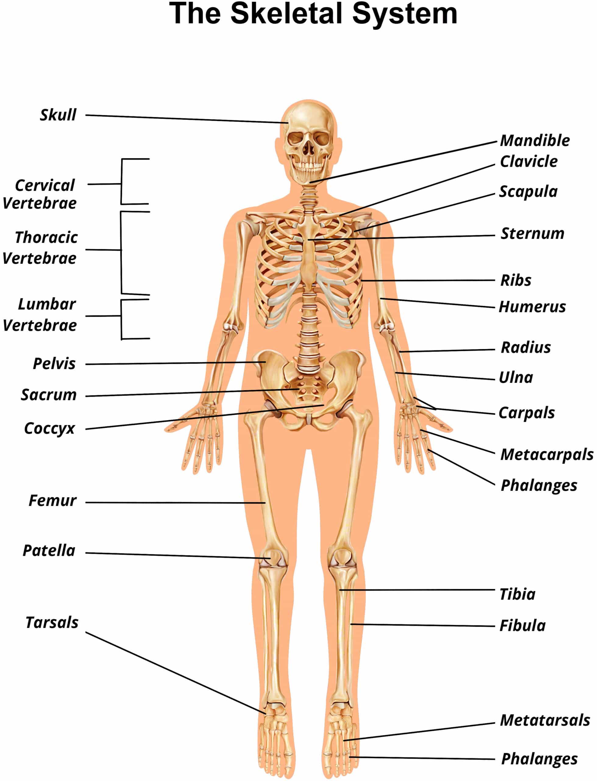
Human infants are born with approximately three hundred bones. As each person grows and develops, that number reduces due to bone fusion. Once a person reaches adulthood, their total number of bones drops down to two hundred and six.1
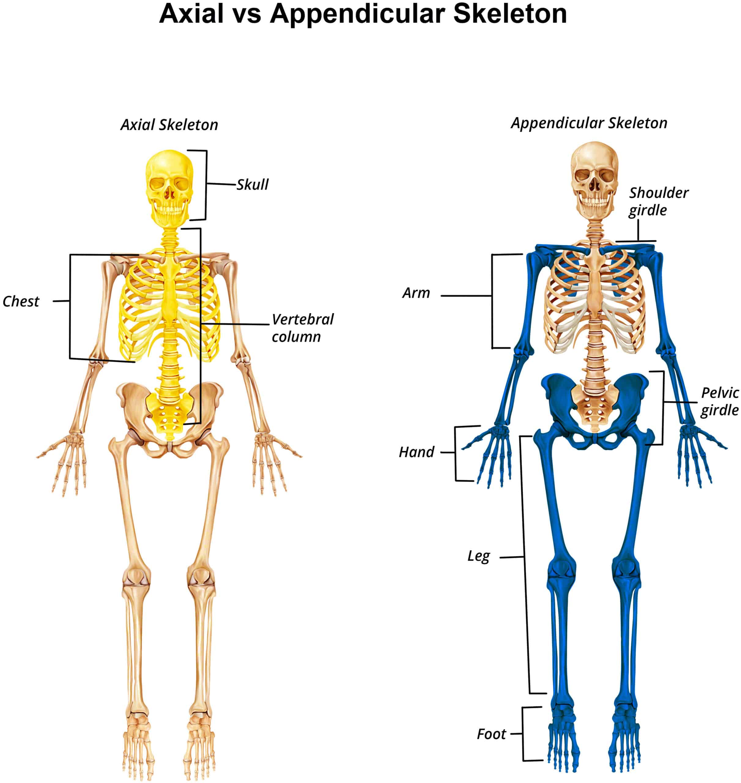
The human skeleton divides into two main parts – the axial and the appendicular skeleton.
The axial skeleton contains central bones such as the:
- Vertebrae
- Ribs
- Sternum
- Skull
It makes up eighty bones in all. The axial skeleton’s primary function is to protect organs in the body’s systems. These bones also contain attachment sites for muscles that assist with global support and balance.
The appendicular skeleton is made of one hundred twenty six bones that create the upper and lower limbs. These bones also include those of the pelvis and the shoulders. Their main function is to support movement in the extremities and serve as the attachment site for muscles.1
Bone Types
Bones are separated into categories based on their appearance. The four main types of bone are:
- Long bones
- Short bones
- Flat bones
- Irregular bones
The names used reflect the appearance of each bone category.3
Surrounding every bone is a tissue called periosteum, which covers most of the bone except for the locations where bones connect at a joint.
The periosteum is a membrane made of two layers – an outer layer made of connective tissue and an inner layer that contains bone stem cells. Blood vessels and nerves run through this membrane.3
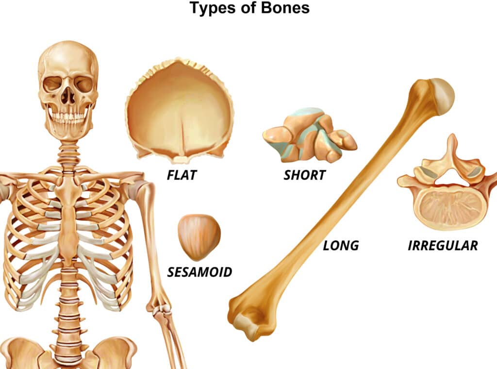
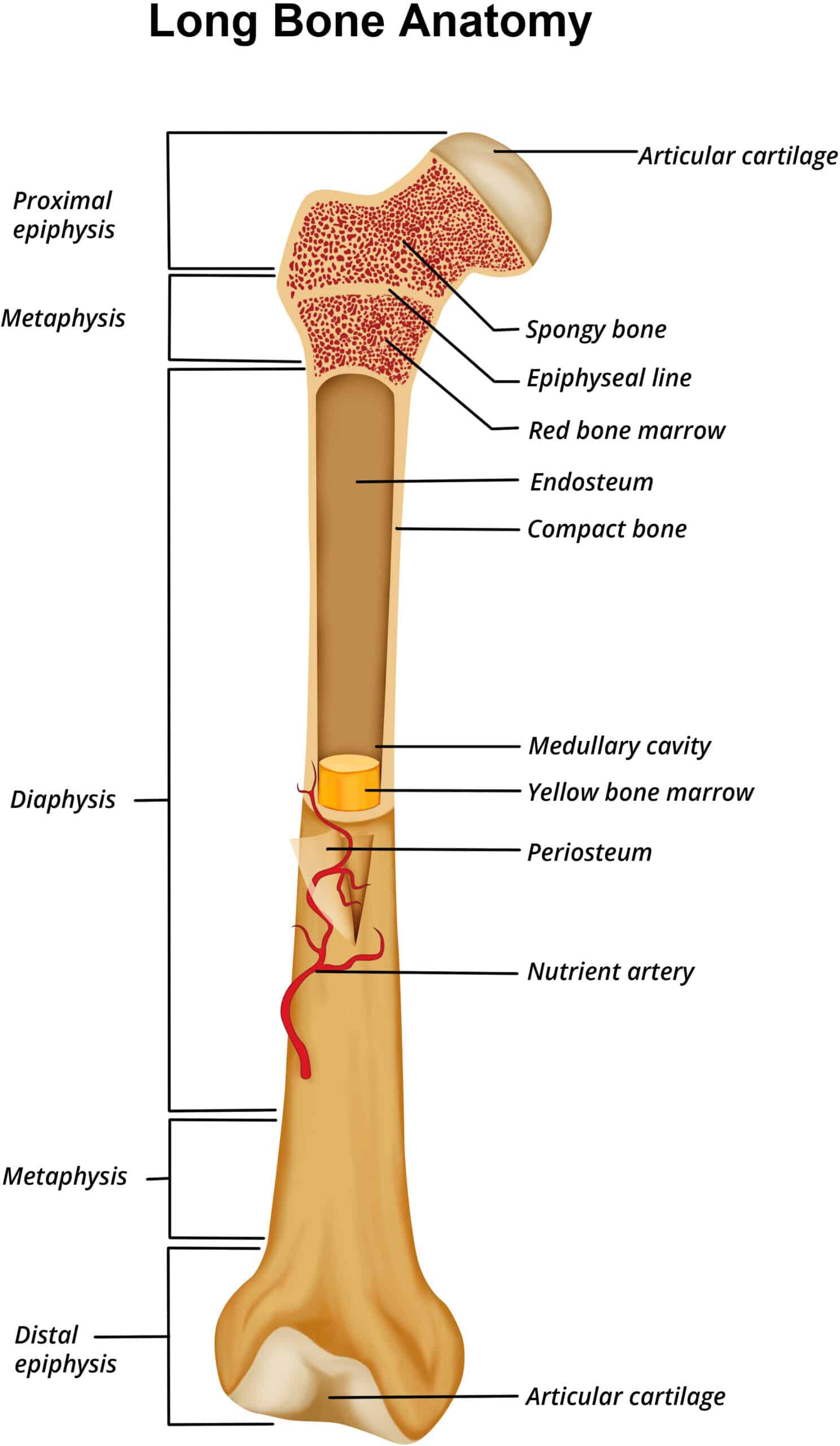
Long Bones
Long bones have a greater length than they do width or height. Most of the limb bones are long bones.
Each long bone has a diaphysis. The diaphysis is a shaft that makes up the middle length of the long bone.
On each side of the diaphysis sits the epiphysis. The epiphysis is the end of the long bone, so each long bone has two epiphyses.3
During childhood, long bones contribute to height increase and limb lengthening through epiphyseal plates. These growth plates are located in between the diaphysis and epiphysis and are made of cartilage that is replaced by bone as the plate grows. They allow for the lengthening of the long bones. Once a person reaches their maximal height and limb lengths in adulthood, the epiphyseal plates become epiphyseal lines that no longer actively contribute to the lengthening of the bones.3
Examples:
- Phalanges in the fingers
- Ulna and radius in the forearm
- Humerus in the arm3
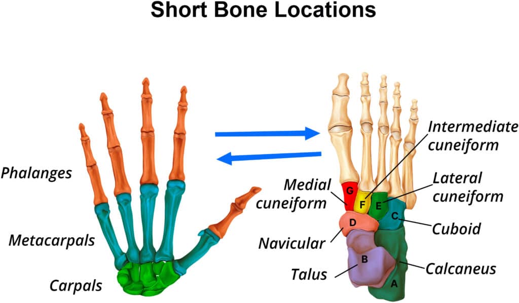
Short Bones
Short bones are cuboid in shape. Their measurements run similarly in length, height, and width.
Examples:
- Wrist bones
- Ankles
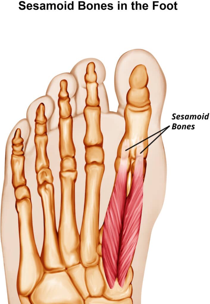
Sesamoid Bones
Sesamoid bones are short bones specifically found in tendons. Many of the sesamoid bones vary in size and location from person to person. They even range in number from person to person.3
Example:
- Patella in the kneecap
Flat Bones
Flat bones are relatively flat as their name implies. Sometimes they have a curve to them, such as in the ribs.
Examples:
- Scapulae in the shoulders
- Cranial bones in the skull:
- Frontal
- Parietal
- Occipital
Temporal3
Irregular Bones
Irregular bones are bones that do not fit into the category of long, short or flat bones. They have varying shapes depending on their specific function.
Examples:
- Cervical vertebrae
- Thoracic vertebrae
- Lumbar vertebrae3
Bone Markings
In addition to categorizing individual bones, scientists also categorize the types of markings found on bones.
There are three main kinds of bone markings:
- Depressions
- Projections
- Surfaces
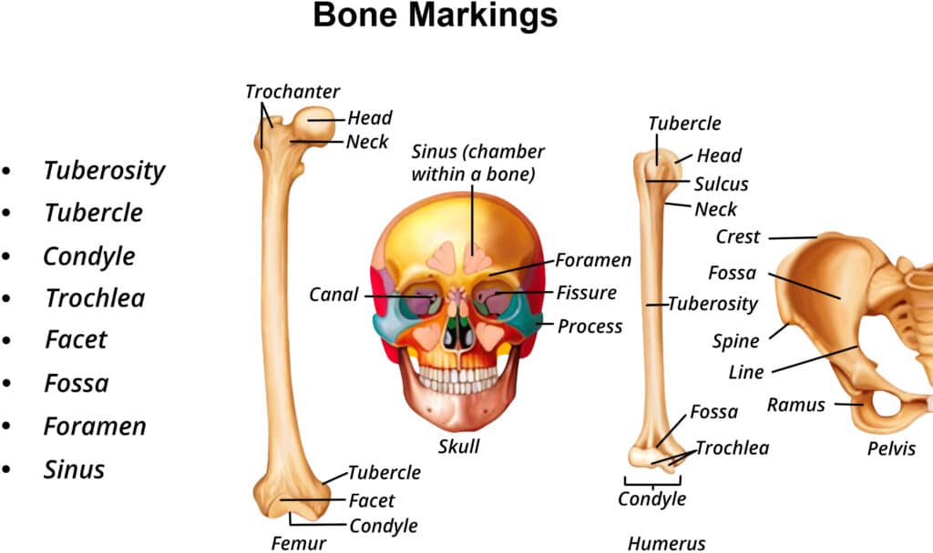
Depressions
Depressions are spaces on the bone through which nerves or blood vessels run through.
These are further divided into categories such as:
- Foramen – holes or openings for nerves and blood vessels to pass through
- Fissure – open slits, grooves, or depressions typically housing nerves and blood vessels
- Fossa – broad and shallow depressions in the bone surface
- Sulcus – a furrow or fissure that in the case of bones typically traces the length of nerves or vessels–also called grooves when specifically discussing sulci on bones
Projections
Projections are the attachment sites of tendon and ligament to bone. Categories of projections include:
- Processes
- Condyle
- Epicondyle
- Process
Surfaces
Surfaces are the parts of the bone that attach to another bone to make a joint. These include facets and heads. Many depressions, projections, and surfaces found on bones have standard names anatomists use to recognize their location in the body.3
Bone Tissue Creation
Bone tissue is composed of many types of cells, including some that are specialized for the skeletal system. Two essential cell types found in the bone tissue are osteoblasts and osteoclasts.
Osteoblasts are cells that help build bone.
Osteoclasts help break down bone.
These two cell types are important, because the body needs to build and break down bones throughout its lifetime.1
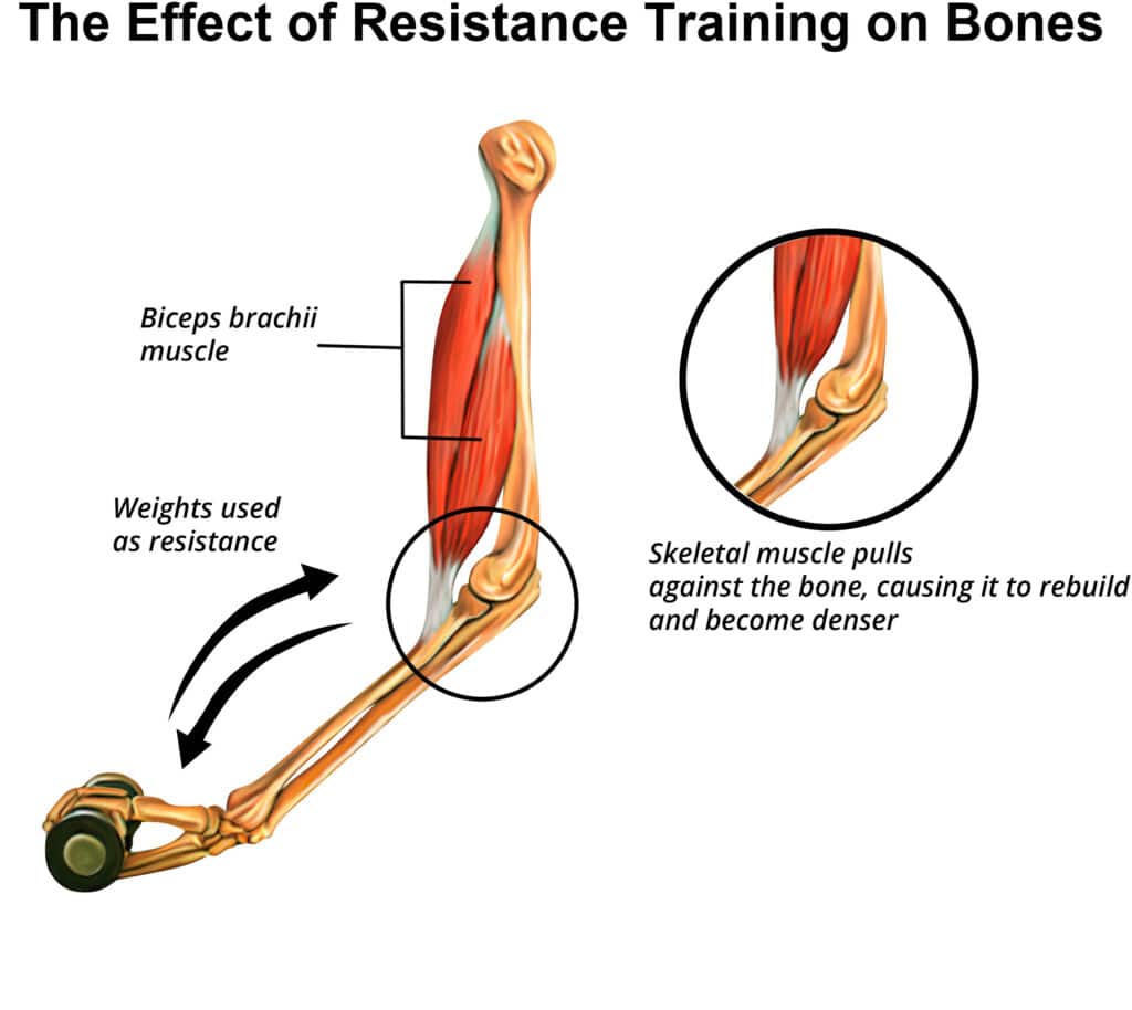
Wolff’s Law
Wolff’s Law is an important theory of the skeletal system related to bone creation.
This theory states that bone adaptations occur in response to the outside environment and the external forces the body experiences.
According to Wolff’s law, bone tissue changes over time by being built or broken down into different shapes, structures, and densities based on the environmental stresses placed on the bone.
For example, someone who becomes sedentary after trading their dog walking career for a new office job now primarily spends their day sitting at a desk. Since the bones now put much less stress on their bones, they begin to be broken down by osteoclasts.
The body does not need as much bone to continue to protect all the organ systems and move. Therefore, over time, this individual’s bones will become thinner and smaller.2
For another example, consider a professional soccer player who spends her day training for her sport. She strength trains at her gym, practices agility with her personal trainer, and does practice drills with her team.
These environmental stressors trigger the skeletal system to react as her body is constantly supporting itself against many and varied outside forces.
Osteoblasts in the bone tissue are busy at work building stronger, denser bones. She needs these strong dense bones to protect her organs as she runs across the soccer field and to support powerful movements like kicking the ball into the goal.2
Connective Tissue in the Skeletal System
While bone tissue makes up a lot of the skeletal system, ligaments and tendons are also part of this body system.
Ligaments and tendons are made of connective tissue rather than bone tissue. Connective tissue differs from bone tissue in that it is more elastic and flexible. It primarily consists of type I collagen fibers surrounded by a mesh of loose connective tissue.4 This allows for mobility and range of motion at each joint.1
Ligaments
Ligaments are connective tissues that connect one bone to another bone. They aid in holding the bones together while allowing a range of motion between the two bones.
Tendons
Tendons are connective tissues that attach muscle to bone, allowing the muscle to stretch without immediately tearing.1
Joints
Non-synovial joints are limited in mobility or completely immobile. They serve to connect bones of protection. The sutures of the skull bones (these bones protect the brain) and the fused vertebrae of the lower spine (these bones protect the spinal cord) are examples of non-synovial joints.1
Synovial Joints
In exercise science and personal fitness training, synovial joints are the main type of joint to focus on, as virtually all movement in the human body occurs around synovial joints.
Synovial joints are also the most common type of joints found in the body. They allow for large range of motion while protecting the bones that make up the joint. Between the bones at this type of joint, is a synovial capsule filled with fluid, conveniently named synovial fluid.
This capsule cushions the bones, preventing potential injury from two bones rubbing directly against each other. In addition to the synovial capsule, ligaments are also often found at synovial joints for added stability of the joint.1
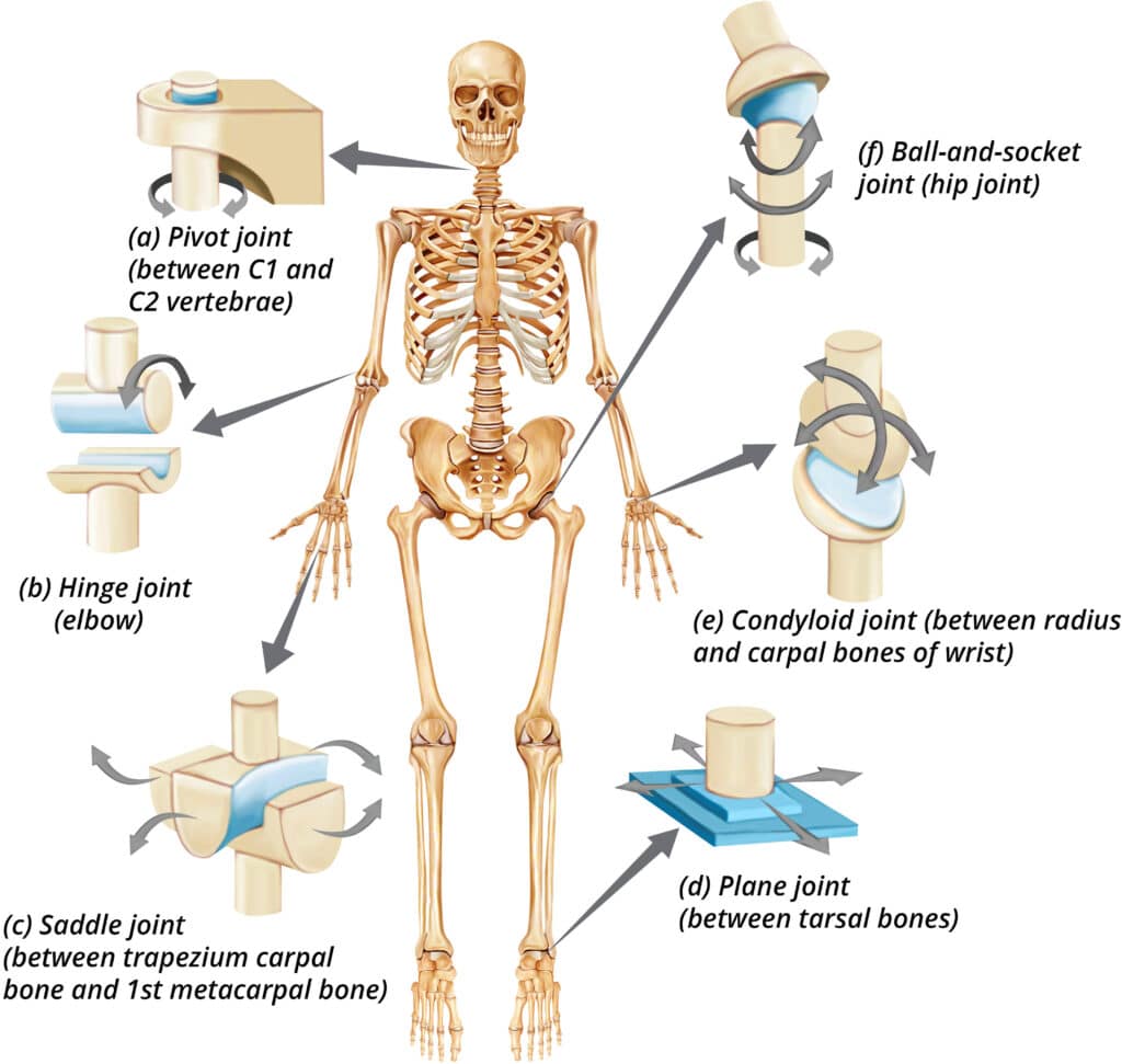
Synovial joints have many different structures allowing for many different functions. Therefore, it makes sense to classify the synovial joints into subtypes based on their structures and functions.
The subtypes of synovial joints are:
- Gliding joints
- Ball-and-socket joints
- Pivot joints
- Hinge joints
- Saddle joints
- Condylar joints1,2,3
Gliding Joints
These are also known as plane joints. They move using a gliding movement as the name suggests – two bones are “gliding” against each other. They only allow for movement in one axis. This type of joint is found in the wrists and in ankles.3
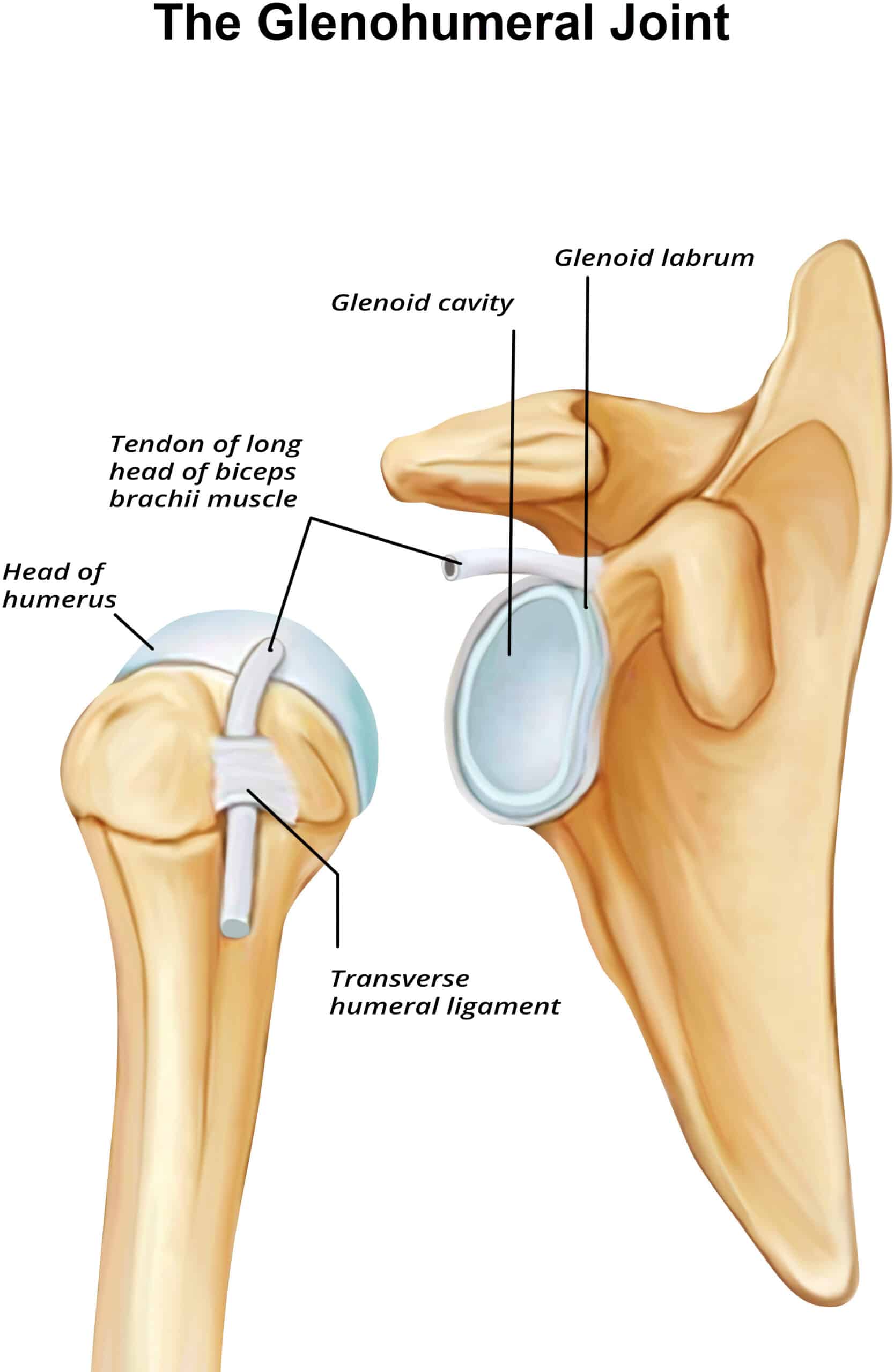
Ball-and-Socket Joints
Ball and socket joints have movements in many axes.
Ball-and-socket joints have lots of mobility but contain much less stability. These joints are often the location of injury due to this lack of stability. Ball-and-socket joints include those in the hip joint and shoulder joints.3
Pivot Joints
Pivot joints create rotating movements. Like ball-and-socket joints, there are only a few joints in the body that display this kind of movement.
The joints in the forearm composed of the ulna and radius are an example. When one places the palm of their hand on a surface in front of them and then flips the hand over so they can see their own palm, they are performing a movement using the pivot joints in their forearm.
Hinge Joints
Hinge joints create a hinge movement – think of a hinge that connects a door to a doorway. They only allow for movement in one axis. Examples of these joints include the elbows, fingers, and toes.3
Saddle Joints
Saddle joints allow for movements in two axes. The joint is named because it looks like a saddle on a horse, with one bone surface surrounding the other. An example of a saddle joint is the saddle joint found in the thumb allowing for special opposable movements.3
Condyloid Joints
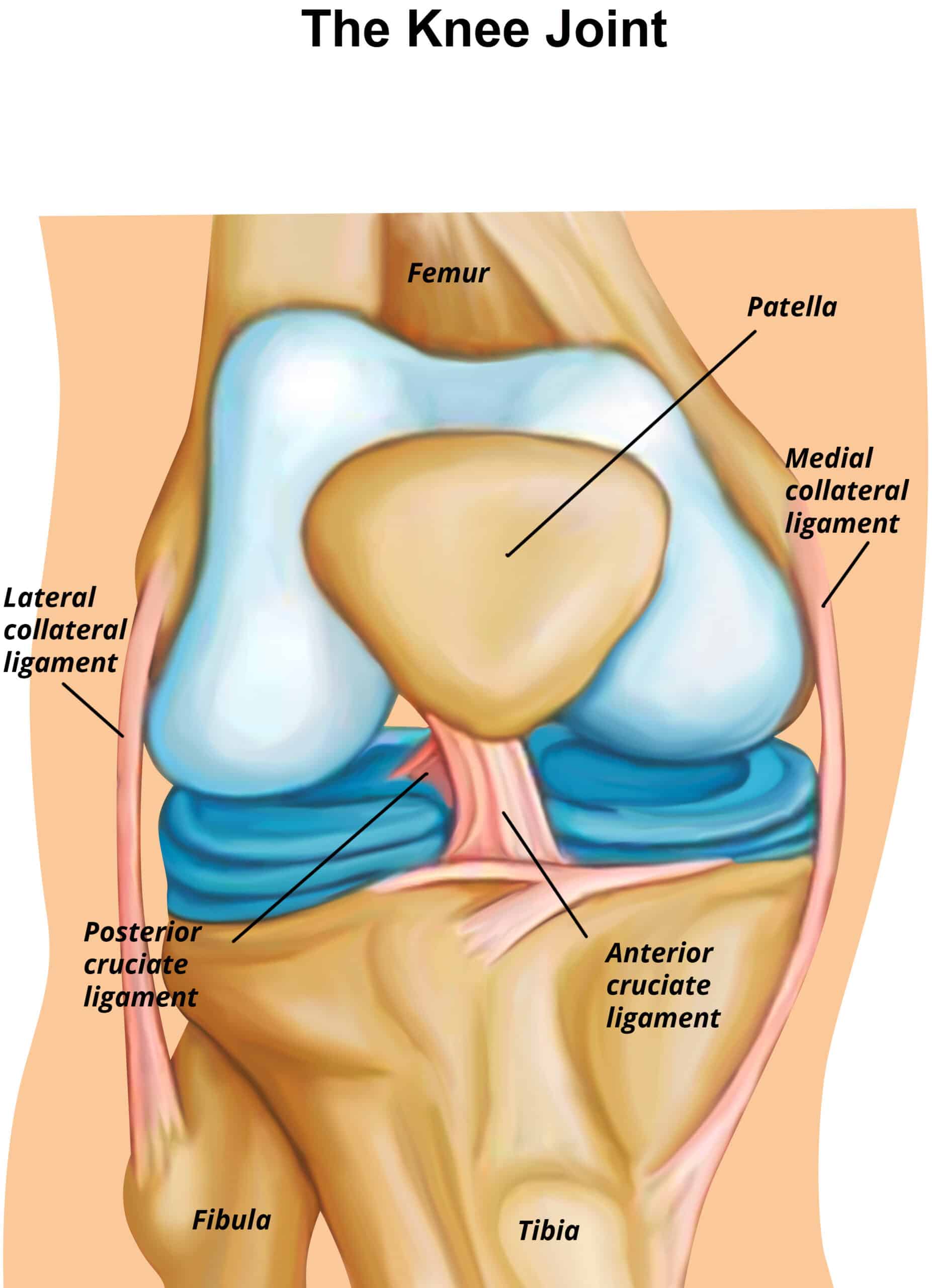
Condyloid joints also allow for movements in two axes. The easiest way to differentiate condylar joints from saddle joints is by their appearance. While saddle joints are shaped like saddles, condyloid joints look like two connected ovals. Each bone surface contributes an oval shape to this joint. Examples of condyloid joints include the joints in the knuckles, some joints in the wrist, and the knee joint.3
Bone and Joint Disease
The skeletal system can be affected by disease like every other body system.
Some of the main bone and joint diseases relevant to personal training are:
- Osteoporosis
- Arthritis
- Osteoarthritis
- Rheumatoid arthritis
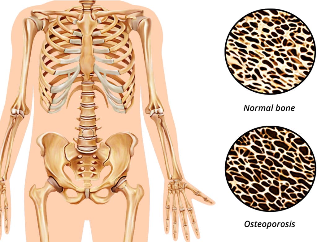
Osteoporosis
Osteoporosis is a disease that causes bone mass to become critically low, increasing the risk of bone breaks. Osteoporosis happens more commonly in women post-menopause, although it can affect anyone at any age.
Proper nutrition can assist in the prevention of osteoporosis. This includes intaking enough dietary calcium under the supervision of a registered dietician, and building bone through weight bearing exercise.
Treatment for osteoporosis includes both medications and weight-bearing exercise to rebuild some of the lost bone.1
Arthritis
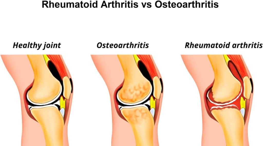
Arthritis is a common disease found in the joints characterized by pain and inflammation at the location of each affected joint. There are two kinds of arthritis commonly encountered on the gym floor: osteoarthritis and rheumatoid arthritis. Both diseases cause damage to the joint.1
Osteoarthritis
Osteoarthritis is the more widespread form of the disease and more frequently seen in older populations; however, individuals of any age are susceptible.
Cartilage at the affected joint site has worn down leading to bone injury at the surfaces of the bones.
Osteoarthritis is treated with medication and exercise.
However, osteoarthritis pain can sometimes increase with sustained activity, so exercise should cease the moment if it becomes too painful.
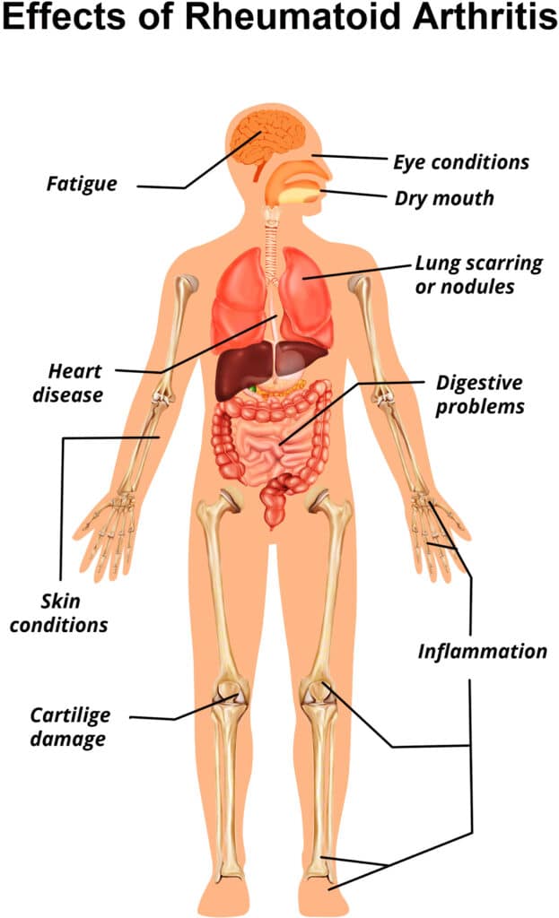
Rheumatoid Arthritis
Rheumatoid arthritis is an autoimmune disease where the body’s immune system attacks the joints. It can affect anyone at any age, but disproportionately affects women.
Rheumatoid arthritis is also treated with medication and exercise. Typically, rheumatoid arthritis pain lessens with sustained activity.
Exercise and the Skeletal System
The theory of Wolff’s Law applies to exercise as it does any other external stressor. Any weight-bearing exercise adds outside stress to the body. Therefore, progressing with any form of weight-bearing exercise will increase bone mass.
Cardiovascular exercise such as running or walking also adds outside stress to the body and can increase bone mass when performed consistently over time.
Strength training is one of the most effective stimuli to increase bone mass due to the ability to load the axial spine very effectively.
The degree of bone mass increase will vary based on the type of exercise and how much stress it places on the bones.3 Any weight-bearing exercise helps prevent osteoporosis and bone breaks, especially as clients age. The denser the bones, the more difficult it is for bones to break.1,3
Summary
The adult skeletal system consists of 206 bones of various sizes, shapes, and structures that support and protect the body. The system is divided into the axial and appendicular skeleton.1
The main types of bones are long bones, short bones, flat bones, and irregular bones. Each of these bones has markings that include depressions, projections, and surfaces.3
Each bone is covered by periosteum. Long bones contain a diaphysis that separates two epiphyses.3 All bones adapt to outside stressors through the work of osteoblasts and osteoclasts as described by Wolff’s Law.1
Tendons and ligaments also help make up the skeletal system. Joints made of two or more bones (and sometimes ligaments) connect the skeletal system and allow for range of motion.
Non-synovial joints help protect organs while synovial joints such as gliding, ball–and–socket, pivot, hinge, saddle, and condylar joints allow for movement.1,2,3
Diseases affecting the skeletal system include osteoporosis, osteoarthritis, and rheumatoid arthritis.1
As predicted by Wolff’s Law, the skeletal system will increase bone mass as an adaptation to many different types of exercise, since exercise causes external stress on the body.3
Improvements to bone mass and the subsequent ability of bones to withstand more force before breaking are key benefits of exercise that occur within the skeletal system in response to training with external resistance.
References
- Sayler, Mary Harwell. The Encyclopedia of the Muscle and Skeletal Systems and Disorders. Facts on File, Inc; 2005.
- Anderson DM, Novak PD, Jefferson K, Elliott MA, eds. Dorland’s Illustrated Medical Dictionary. 30th ed. Philadelphia, PA. Elsevier; 2003.
- Marieb, R.N., Ph.D., Elaine N., Hoehn, M.D., Ph.D., Katja. Human Anatomy and Physiology. 11th ed. Pearson; 2019.
- Zschäbitz A. Anatomie und Verhalten von Sehnen und Bändern [Structure and behavior of tendons and ligaments]. Orthopade. 2005;34(6):516-525. https://doi.org/10.1007/s00132-005-0799-4